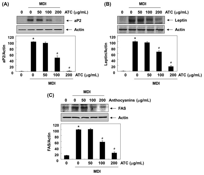
Fig. 6.
Effects of anthocyanins on the levels of adipocyte expressed genes expression in the differentiated 3T3-L1 cells. The proteins were isolated cells grown under the same conditions (as shown in Fig. 2), and the cellular proteins were separated electrophoretically using SDS-polyacrylamide gels, and transferred onto membranes. The membranes were probed with the indicated antibodies. The proteins were visualized using an ECL detection system. Actin was used as an internal control. The relative ratios of the expression in the results of Western blotting are presented at the bottom of each result as the relative values of the expressions in the MDI-treated group. The data were expressed as the mean ± SD of three independent experiments (*p < 0.05, vs. undifferentiated control; #p < 0.05, vs. differentiated control).

