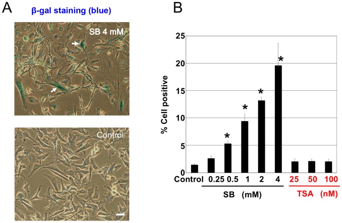
Figure 2.
Effect of SB and the HDAC inhibitor TSA on SA β-gal staining. (A) A172 cells treated with SB (0–4 mM) or TSA (25–100 nM) for 4 days were stained with SA β-gal. β-gal-positive cells are indicated by white arrows (scale bar=20 µm). (B) β-gal-positive cells in (A) were analyzed and counted. Results are presented as the mean ± standard deviation (n=4); *P<0.01 vs. control. SA β-gal, senescence-associated β-galactosidase; SB, sodium butyrate; HDAC, histone deacetylase; TSA, trichostatin A.

