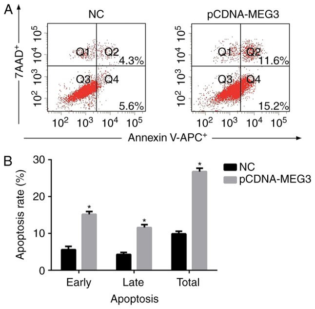
Figure 2.

The effect of MEG3 on MG63 cells apoptosis. (A) After transfected MG63 cells for 48 h, cells were collected and stained with Annexin V-APC/7AAD and then examined by flow cytometry. The early apoptosis rate (Q4) of the NC group and pCDNA-MEG3 group were 5.61±0.92% and 15.19±0.66%, respectively. The late apoptosis rate (Q2) was 4.26±0.52% in the NC group and 11.64±0.79% in the pCDNA-MEG3 group. (B) The histogram showed the apoptosis rate obtained from NC and pCDNA-MEG3 group. The total apoptosis rate of the pCDNA-MEG3 group was higher compared with the NC group (*P<0.05).
