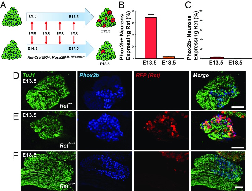
Fig. 1.
Ret is highly expressed in chemosensory geniculate neurons early in development. (A) Experimental strategy for tracing Ret expression in embryonic geniculate ganglia. Tamoxifen was administered to Ret-Cre/ERT2; Rosa26LSL-TdTomato/+ reporter mice at E9.5 to E12.5 with E13.5 analysis (Upper) and E14.5 to E17.5 with E18.5 analysis (Lower). (B) Quantification of the proportion of chemosensory (Phox2b+) neurons expressing Ret demonstrates widespread expression within chemosensory neurons (E13.5; n = 3), but Ret expression is extinguished perinatally (E18.5; n = 4). (C) Quantification of the proportion of somatosensory (Phox2b−) neurons expressing Ret. (D–F) Immunofluorescence with TuJ1 (green), Phox2b (blue), and RFP (indicating Ret; red) with merged images (Right). (D) Staining in a Ret+/+ littermate demonstrates the specificity of the RFP antibody. (E and F) Ret was widely expressed in chemosensory neurons at the E13.5 analysis time point (E) but largely absent upon analysis at E18.5 (F). Note that the TG in the upper right hand corner of F has many Ret+ neurons at E18.5. Error bars indicate mean ± SEM. (Scale bars, 50 µm.)

