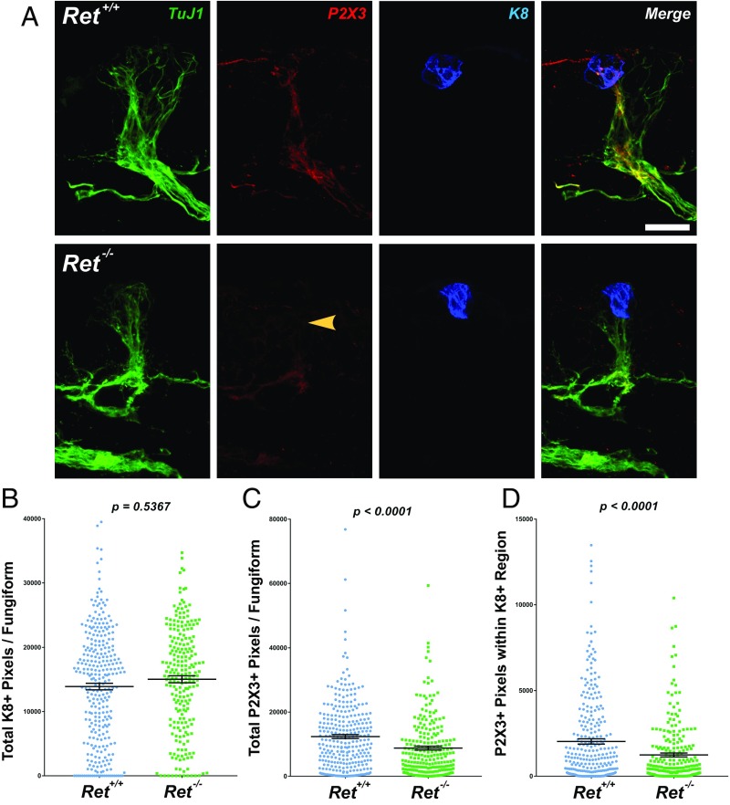
Fig. 4.
Loss of Ret results in deficits in fungiform papilla chemosensory innervation. (A) Tongues were collected from E18.5 Ret+/+ and Ret−/− mice, serially sectioned at 50 µm, and immunostained for TuJ1 (green), P2X3 (red), and K8 (blue) and merged. Many more examples of FPs lacking apically projecting P2X3+ fibers were observed in Ret−/− compared with Ret+/+ tongues (yellow arrowhead). (B) All fungiform papillae from n = 4 mice were imaged (n = 300 Ret+/+ FPs and n = 230 Ret−/− FPs) and quantified as described in Experimental Procedures. When analyzing the entire papilla, no differences were observed in the number of K8+ pixels per FP (P = 0.5367). (C) A highly significant reduction in P2X3+ pixels was observed in Ret−/− mice (P < 0.0001). (D) When analyzing only the nerve fibers present within the K8+ region, P2X3+ pixels were substantially reduced (P < 0.0001). The graphs in B–D display individual data points (colored circles and squares), while the mean ± SEM is indicated by the black lines. (Scale bar, 25 µm.)

