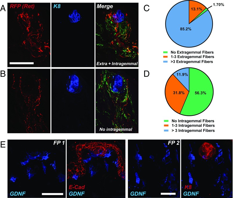
Fig. 6.
Distribution pattern of Ret+ nerve fibers and GDNF+ cells within fungiform papillae. (A) Whole tongues from adult Ret-Cre/ERT2; Rosa26LSL-TdTomato/+ mice were immunostained for RFP (indicates Ret; red), K8 (blue), and TuJ1 (green; present in merged image). One hundred seventy-six FPs from n = 5 individual mice were imaged and the pattern of Ret expression was categorized as intragemmal (within the K8+ region) or extragemmal (outside the K8+ region). This is an example of an FP with extensive (>3 fibers) intragemmal and extragemmal innervation by Ret+ fibers. (B) Example of an FP with extensive (>3 fibers) extragemmal labeling but no intragemmal Ret+ fibers. (C) Quantification of the innervation density of Ret+ extragemmal fibers indicates that the vast majority of FPs have extragemmal fibers present (98.3%), most of which (85.2%) have greater than three fibers. (D) Quantification of the innervation density of intragemmal Ret+ fibers demonstrates that many FPs (43.7%) have an intragemmal projection pattern. (E) Five daily doses of TMX were administered to adult GDNF-IRES-Cre/ERT2; Rosa26LSL-TdTomato/+ via i.p. injection. Tongues were then fixed, preserved, serially sectioned, and immunostained for RFP (indicating GDNF; blue) and E-cadherin (red; Left, FP 1) or K8 (red; Right, FP 2). All FPs were imaged from n = 4 individual mice. We observed a variable pattern of expression of GDNF within the TB region (ranging from 0 to 2 cells generally present within the basal aspect of the TB) but strong labeling of GDNF in the perigemmal space immediately surrounding the TB. In addition, GDNF+ cells were observed in the E-Cad+ trenches at the base of the FP. On occasion, GDNF+ cells were observed in the mesenchymal core of the FP. (Scale bars, 50 µm.) In all cases, Cre-negative littermate controls were always utilized to control for RFP immunostaining specificity.

