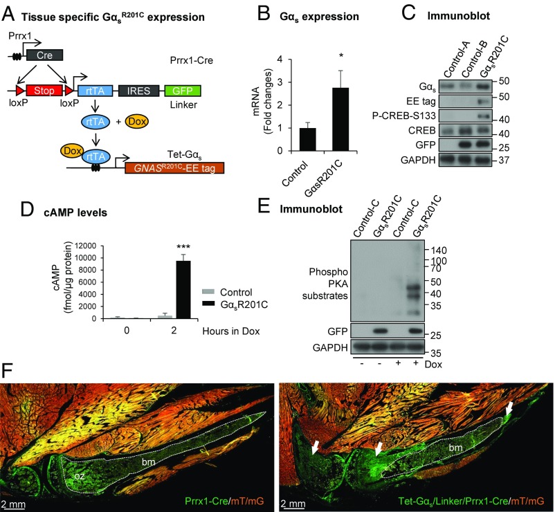
Fig. 1.
Generation of the transgenic mouse model of fibrous dysplasia (FD). (A) Schematic representation of the transgenic mouse model to express Gαs-activating mutations in a tissue-specific manner. (B) Quantitative PCR (qPCR) analysis (n = 6) and (C) immunoblotting showing elevated expression of Gαs in limb bone from GαsR201C mice relative to control after doxycycline (Dox) administration. Both intercellular cyclic adenosine monophosphate (cAMP) level (D) and protein kinase A (PKA) phosphorylation (E) are dramatically elevated in GαsR201C BMSCs in response of Dox treatment. (F) Fluorescence imaging from Rosa26 mT/mG reporter mice (mT/mG) shows the expression of Tet-Gαs/linker/Prrx1-Cre cassette and the initiation of FD (white arrows). White dashed lines outline ossification zone (oz) of growth plate and bone marrow (bm) in control (Prrx1-Cre/mT/mG) tissue or unaffected bone marrow (bm) in mutant (Tet-Gαs/linker/Prrx1-Cre/mT/mG) tissue. The genotype of the mice corresponds to control-A = linker, control-B = linker/Prrx1-Cre, control-C = Tet-Gαs/linker, GαsR201C = Tet-Gαs/linker/Prrx1-Cre. Note: control mice, if not indicated, were selected randomly from mice which did not include the three Tet-Gαs/linker/Prrx1-Cre transgenes. These terms are also applied for the rest of figure legends. Data are presented as mean ± SD, and significance was calculated by Student’s t test (P > 0.05; *P < 0.05; and ***P < 0.001).

