Fig. 3.
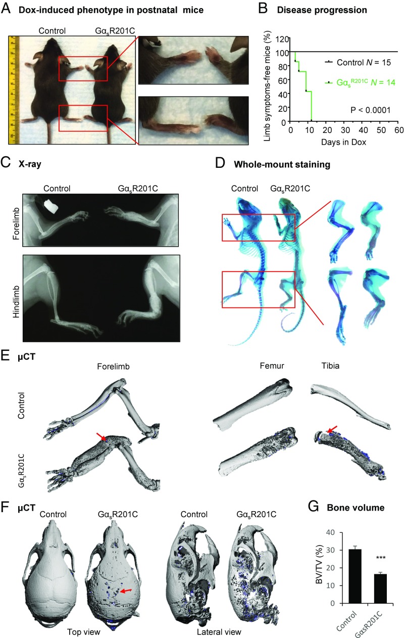
FD lesions in limbs and skulls of postnatal GαsR201C mice. (A) Representative image of the expanded limbs of adult GαsR201C mice. (B) Kaplan–Meier curve of mice free of visible disease symptoms (limb expansion and limping behavior). GαsR201C (n = 14) mice developed bone lesions within 14 d after Dox administration. Control (n = 15) mice remained lesion-free. (C) X-ray examination shows the expansive deformity of long bones. A mixture of lucent and sclerotic images, representing cortical lytic-sclerotic changes, described as ground glass appearance found in human FD patients. (D) Whole-mount staining showing the expansive deformity and lytic changes of the limb bone from GαsR201C mice. (E) μCT examination of limbs showing the lytic-sclerotic lesions with subtle spontaneous fracture (red arrows). (F) μCT examination of skulls, showing the lytic defect (red arrow). (G) Bone volume (BV) fraction (BV/TV) in the regions of limb with FD lesions (total volume, TV) and the corresponding regions in control mice (n = 4). Data are presented as mean ± SD, and significance was calculated by Student’s t test (***P < 0.001).

