Fig. 7.
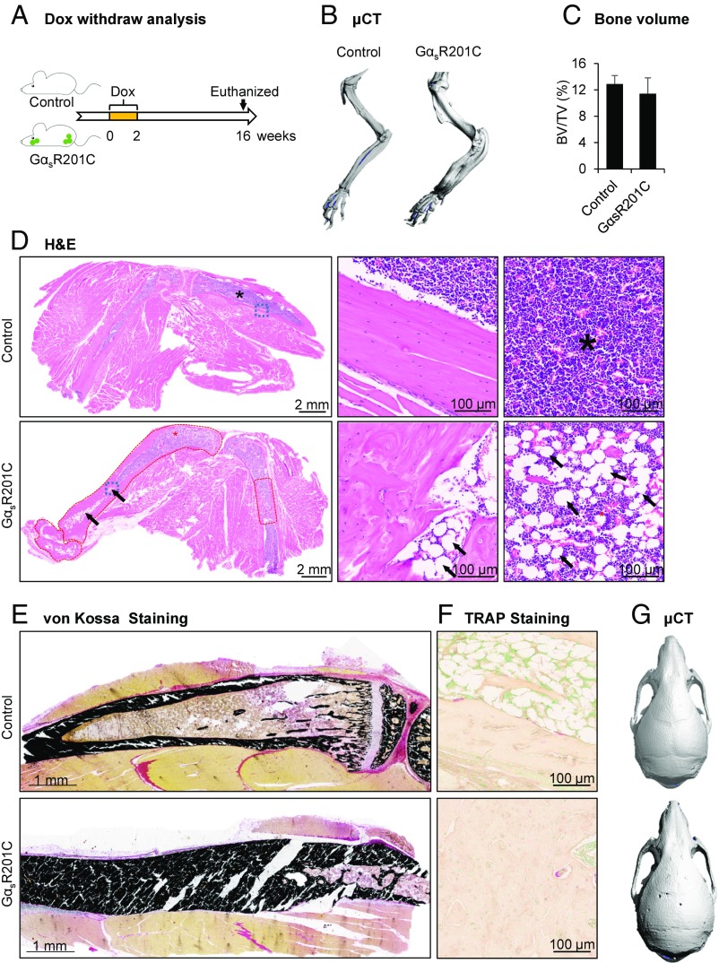
Reducing GNAS expression following Dox withdrawal results in clinical improvement of the preexisting FD bone lesions. (A) Schematic representation of the Dox treatment of control and GαsR201C mice. (B) μCT examination showed the slightly thickened limb bone of the GαsR201C mice without porous-like lytic phenotype or fracture. (C) No significant difference of bone volume fraction between control and GαsR201C mice (n = 4). (D) H&E section of both control and GαsR201C mouse. The preexisting FD lesions circled in red exhibit a dramatic recovery, dense cortical bone with disordered trabecular processes. The diaphyseal marrow cavity exhibits clear boundaries, although slightly narrower than in littermate control, and are composed of hematopoietic cells and adipocytes (black arrows). Details can be better viewed in the Middle and Right which represent higher magnification of the blue squares and stars area in Left, respectively. Note the collagen-rich fibrocellular matrix and multinucleated giant cells are not present at this stage. (E) Von Kossa staining of undecalcified limb tissue shows normal mineralization of cortical bone without lesional osteoid in GαsR201C mice. (F) TRAP staining shows dramatically reduced number of osteoclasts in remineralized bone 16 wk after discontinuing Dox (compare with Fig. 6A). (G) μCT examination of the recovery skull of GαsR201C mice compared with its littermate. Data are presented as mean ± SD, and significance was calculated by Student’s t test. No significant differences were observed.

