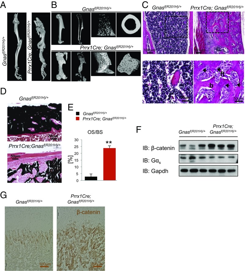
Fig. 2.
Prrx1-Cre; Gnasf(R201H)/+ mutant mice exhibit FD phenotypes. (A and B) Representative µCT scans of forelimbs from P21 mice with the indicated genotypes. Long bones were deformed in the mutant. Longitudinal and cross-sectional images of the humerus (B) showed that bone marrow space was occupied by trabecular bone in mutant mice. (C) Representative H&E staining of the trabecular region of P21 mouse humerus. Boxed regions are enlarged (Bottom); b, bone; ft, fibrous tissues. (D) von Kossa staining showing mineralization in humerus sections of P21 littermate control and mutant mice. (E) Quantification of mineralization in humerus from P21 mice as the percent of osteoid surface (OS) of bone surface (BS) (n = 3). (F) Western blot analysis for β-catenin and Gαs from the cell lysates of P6 humerus. IB, immunoblotting. (G) Immunohistochemistry of β-catenin on humerus sections of P6 littermate control and mutant mice. **P < 0.001; data are presented as mean ± SD.

