Fig. 3.
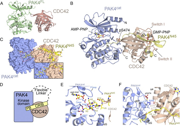
Overall structure of PAK4 in complex with CDC42. (A) Ribbon diagram showing the structure of PAK4FL-CDC42. (B) Ribbon diagram showing the ternary structure of PAK4cat-PAK4N45-CDC42. AMP-PNP and GMP-PNP indicate nucleotide analog. (C) Same orientation as B showing the surfaces of PAK4cat and CDC42. PAK4N45 is shown in stick format. Location of the inset is shown with a box. (Inset) A rotation and zoom of the region boxed in red. (D) Schematic of the interaction. PB indicates polybasic region. (E) Close-up of interaction between the N terminus of PAK4N45 and the PAK4 substrate binding site. The β and γ phosphates of AMP-PNP are not visible in the electron density. (F) Close-up of the interaction between CDC42 and PAK4 showing the extended interface. Secondary structure elements are labeled.

