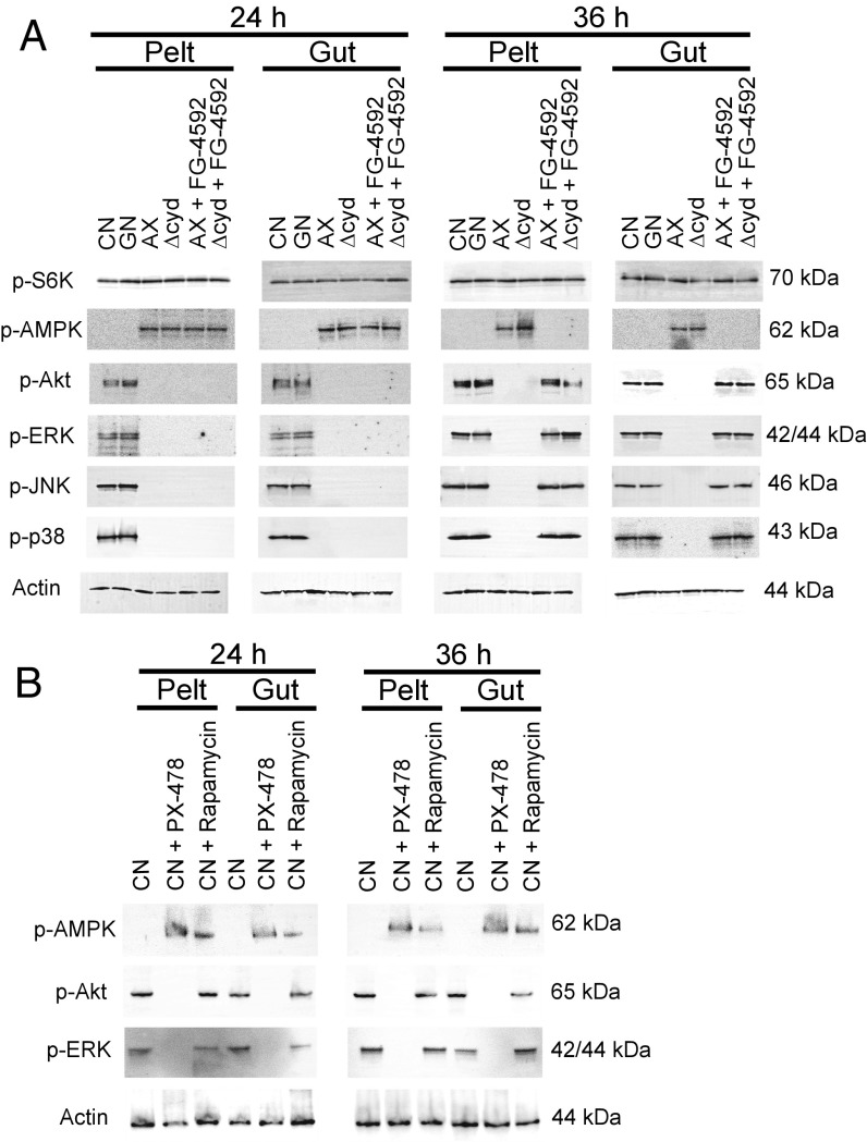
Fig. 2.
Gut bacteria and pharmacological manipulation of HIF-α affect the phosphorylation status of proteins in the TOR, AMPK, and IIS pathways plus select other MAPKs. (A) Immunoblots of pelt and gut extracts from conventional (CN) larvae, gnotobiotic larvae inoculated with wild-type E. coli (GN), axenic larvae (AX), gnotobiotic larvae inoculated with ΔcydB-ΔcydD::kan E. coli (Δcyd), AX with FG-4592 (12 h posthatching), or Δcyd treated with FG-4592 (12 h posthatching). Samples were collected at 24 h or 36 h and probed with antibodies to p-S6K, p-AMPK, p-Akt, p-ERK, p-JNK, p-38, or actin, with molecular masses of each protein target indicated to the right. (B) Immunoblots of pelt and gut extracts from CN larvae and CN larvae treated with PX-478 (12 h posthatching) or rapamycin (12 h posthatching). Samples were collected at 24 h and 36 h, and were probed with antibodies to p-AMPK, p-Akt p-ERK, or actin. The molecular masses of each protein are indicated to the right of each blot.

