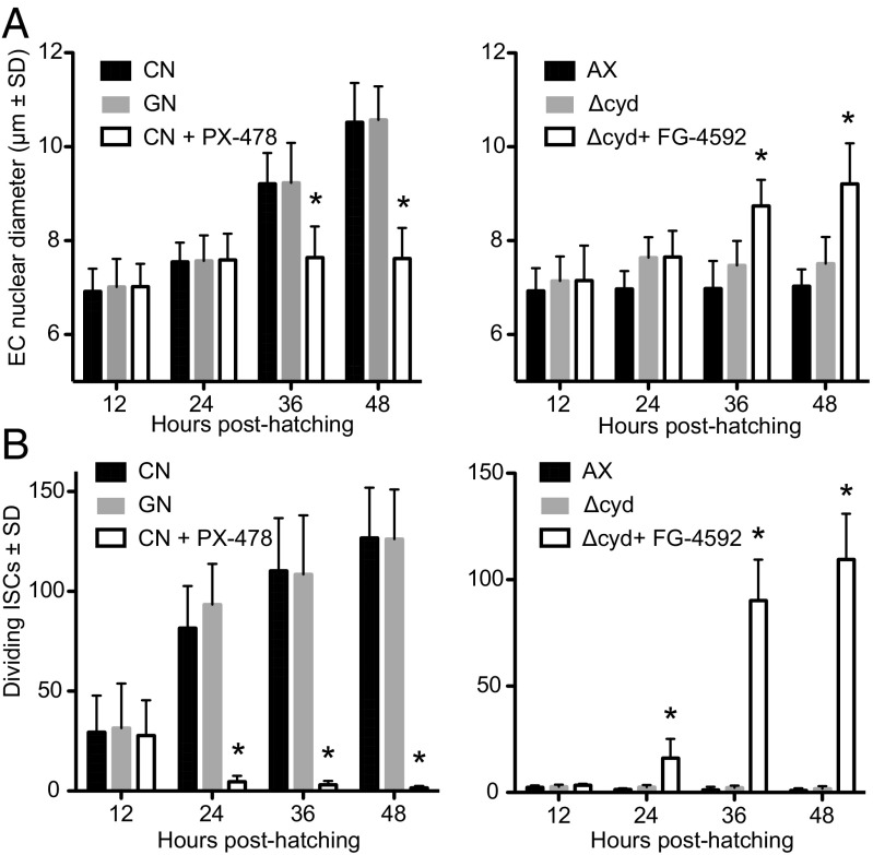
Fig. 4.
Gut bacteria and pharmacological manipulation of HIF-α affect EC size and ISC proliferation. (A) EC size as measured by nuclear diameter in conventional (CN) larvae, gnotobiotic larvae inoculated with wild-type E. coli (GN), or CN larvae treated with PX-478 (Left), and in axenic larvae (AX), gnotobiotic larvae inoculated with ΔcydB-ΔcydD::kan E. coli (Δcyd), or Δcyd treated with FG-4592 (Right). Samples were measured from 12 to 48 h posthatching. An asterisk above a bar at a given time point indicates the treatment significantly differed from that of CN larvae (Left) or Δcyd (Right), which served as controls (analysis at each time point by ANOVA followed by a post hoc Dunnett’s test, P < 0.05). (B) Dividing ISCs as measured by PH3 labeling in CN larvae, GN, or CN larvae treated with PX-478 (Left), and AX, Δcyd, or Δcyd treated with FG-4592 (Right). An asterisk above a bar at a given time point indicates the treatment significantly differed from that of CN larvae (Left) or Δcyd (Right) as in A.

