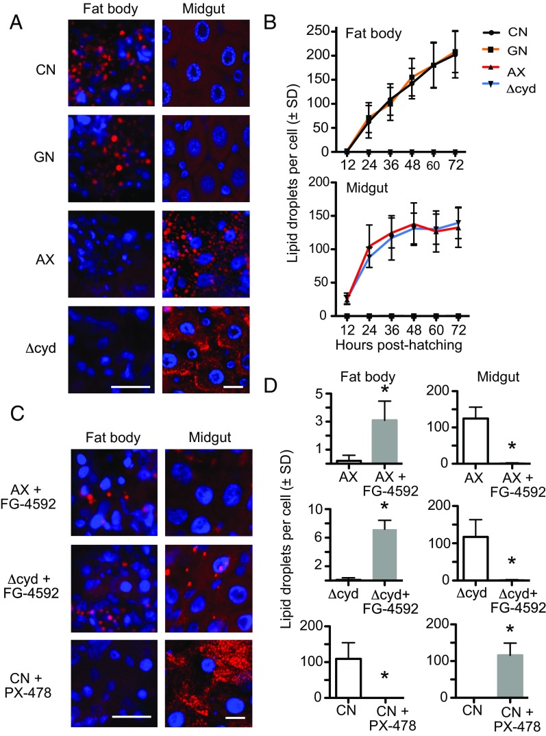
Fig. 5.
Gut bacteria and pharmacological manipulation of HIF-α affect lipid accumulation in the fat body and midgut. (A) Nile red staining of neutral lipids (red) in fat body adipocytes and midgut ECs from conventional (CN) larvae, gnotobiotic larvae inoculated with wild-type E. coli (GN), axenic larvae (AX), and gnotobiotic larvae inoculated with ΔcydB-ΔcydD::kan E. coli (Δcyd) at 36 h posthatching. Cell nuclei are counterstained with Hoechst 33342 (blue). (Scale bars: fat body and midgut images, 20 μm.) (B) Quantification of neutral lipid droplets per fat body adipocyte (Upper) or midgut EC (Lower) from CN larvae, GN, AX, and Δcyd from 0 to 72 h posthatching. For both tissues, CN larvae and GN significantly differed from AX and Δcyd by 24 h posthatching (ANOVA followed by a post hoc Tukey–Kramer honest significant difference test, P < 0.05). (C) Nile red staining of neutral lipids (red) in fat body adipocytes and midgut ECs from AX and Δcyd treated with FG-4592 or CN larvae treated with PX-478 at 36 h posthatching. (Scale bars as in A.) (D) Comparison of neutral lipid droplets per adipocyte or midgut EC in: AX versus AX + FG-4592 treated larvae, Δcyd versus Δcyd + FG-4592 treated larvae, and CN larvae versus CN + PX-478-treated larvae. Larvae were drug-treated at 12 h posthatching, and samples were analyzed at 36 h posthatching. An asterisk above a bar indicates the FG-4592 or PX-478 treatment differed from that of the nontreated control (t test, P < 0.01).

