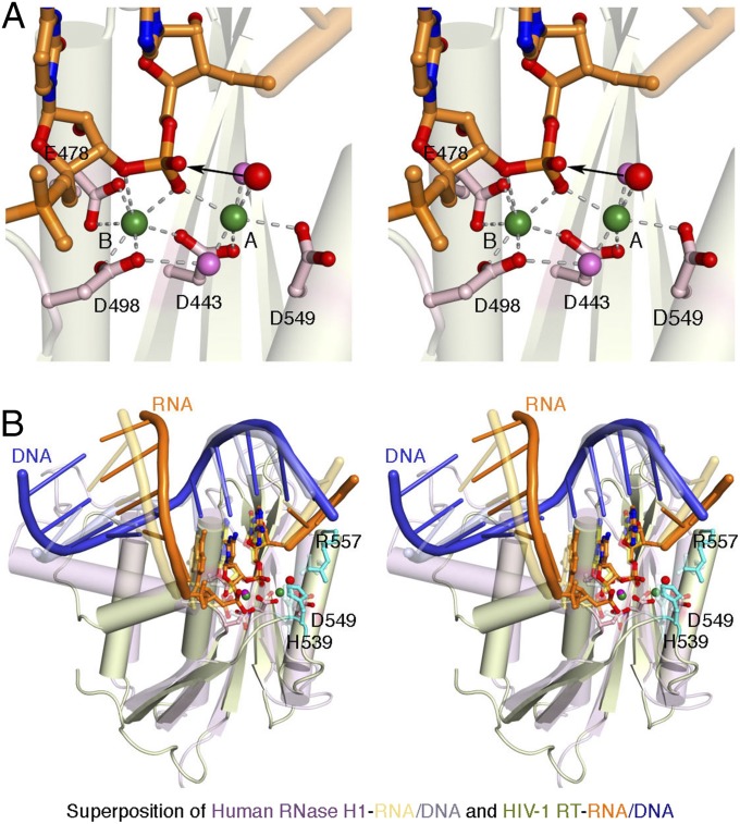
Fig. 2.
The RT-RNA/DNA structure in crystal 1. (A) Closeup stereoview of the RNase H active site. The arrowhead marks the nucleophilic attack of the scissile phosphate. Coordination of the two Ca2+ ions is indicated by the dashed lines. Oxygen atoms are shown in red, nitrogen in blue, and water molecules in pink or red (nucleophile). (B) Stereoview of the superposition of human RNase H1 and HIV-1 RT. The cellular RNase H is shown in pink, with the RNA and DNA in light orange and blue, respectively. The retroviral RNase H is shown in light green, and the RNA/DNA hybrid is in orange and blue. In addition to the catalytic carboxylates, H539 and R557 of RT are shown as sticks.

