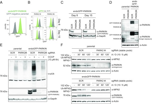
Fig. 1.
endoGFP-PARKIN cells display PARKIN level-dependent pUb accumulation. (A) Clonal JumpIN Cas9+ cells carrying a stable transgene driving GFP-PARKIN expression from 4.5 kb of the endogenous PARK2 promoter (endoGFP-PARKIN; green) or not (GFP-negative parental cells; gray) were assessed for GFP-dependent fluorescence by flow cytometry. (B and C) endoGFP-PARKIN cells display near-complete CRISPR/Cas9-mediated GFP-PARKIN depletion. Cells expressing either an SCR sgRNA or one of two different PARK2-specific sgRNAs (#i, #ii) were assessed for GFP-dependent fluorescence (B) and GFP-PARKIN protein levels (C) 8 or 15 d postinfection. (D) Comparison of GFP-PARKIN protein levels in endoGFP-PARKIN cells to endogenous PARKIN. Protein lysates from stable cell pools expressing either an SCR sgRNA or a PARK2-specific sgRNA (#i) were assessed by WB analysis using indicated antibodies. (E) PARKIN-dependent pUb accumulation in JumpIN cells treated with CCCP. endoGFP-PARKIN cells or parental JumpIN cells expressing PARK2-specific or SCR sgRNAs for 8 d were treated with 10 µM CCCP or vehicle for 6 h, and lysates probed with the antibodies indicated. (F) PARKIN-dependent MFN2 ubiquitination and degradation in parental cells (Upper) or endoGFP-PARKIN cells (Lower). WB analysis using indicated antibodies of stable cell pools expressing PARK2-specific or SCR sgRNAs treated with A/O for the time indicated. WB, Western blot.

