Fig. 6.
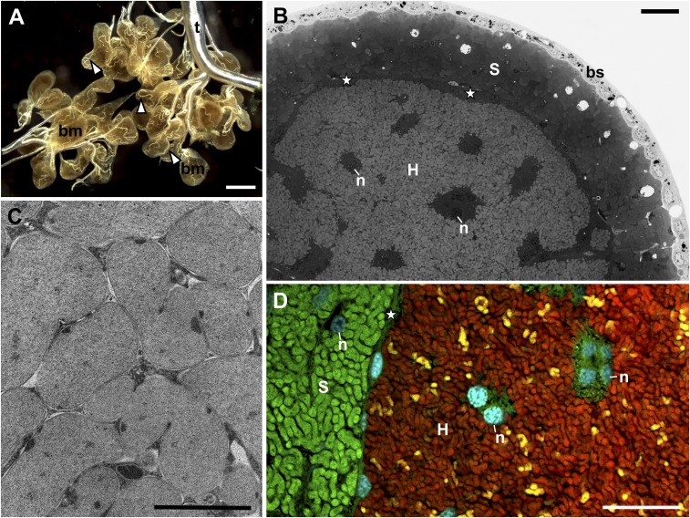
The organization of a cicada bacteriome. (A) The tissue-level view of a dissected male bacteriome from Tettigades sp. 1, which is divided into lobes that are entwined with tracheoles (white arrowheads). bm, bacteriome; t, tracheal system; stereoscope microscope. (Scale bar: 500 μm.) (B) The internal organization of the bacteriome lobe of T. lacertosa, representative of the genus. The external layer consists of simple epithelium (bs, bacteriome sheath). The layer underneath consists of distinct bacteriocyte cells filled with Sulcia (S), and the central part of the bacteriome consists of a single large syncytium filled with cells of Hodgkinia (H). n, nucleus; star, external area of the syncytium that houses Hodgkinia. Light microscope, methylene blue staining. (Scale bar: 10 μm.) (C) Hodgkinia cells in the cytoplasm of the syncytium of Tettigades undata. Note close proximity among cells and their irregular shape. Transmission electron microscope. (Scale bar: 1 μm.) (D) Fluorescence microphotograph of the central part of the bacteriome lobe of T. chilensis specimen TETCHI. Cyan corresponds to the Hoechst, universal DNA stain. Green and red represent the signal of fluorescently labeled probes that target 16S rRNA of Sulcia and Hodgkinia, respectively. Yellow represents the signal of a probe specific to 16S rRNA of only one of Hodgkinia variants, TETCHI2. (Scale bar: 10 μm.) Annotations are the same as in B.

