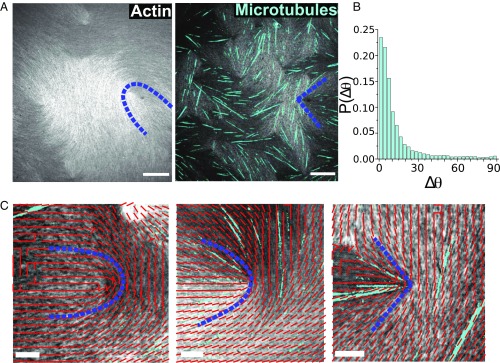
Fig. 3.
Experiments on changing actin-based LC’s elasticity by adding rigid microtubules. (A) Optical images of actin LC without and with microtubules. Blue dashed lines highlight the change in defect shape from U to V. (Scale bar, 30 m.) (B) Probability distribution of the angle between microtubule orientations and the local F-actin director fields. (C) Optical images of defects overlaid with the corresponding director field. Microtubule number density increases from left to right followed by the defect shape change from U to V. (Scale bar, 10 m.)

