Fig. 3.
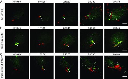
Multiple autophagic processes sequentially target intracellular bacteria. Selected Z-stacked micrographs from time-lapse microscopy experiments of GFP-LC3 (green) BMMs infected with mCherry L. monocytogenes strains (red). (A) GFP-LC3 WT BMMs infected with WT bacteria. Arrowheads show a LC3+ vacuole that was removed from a bacterium and formed a membrane aggregate in the host cytosol. (B) GFP-LC3 WT BMMs infected with the triple mutant. Arrowheads show bacteria that colocalized with LC3. Note that the triple mutant colocalized with LC3 multiple distinct times. (C) GFP-LC3 Fip200−/− BMMs infected with the triple mutant. Arrowheads show a LC3+ vacuole that was removed from bacteria and formed a membrane aggregate in the host cytosol. Times are indicated (h:min:s) above each micrograph. (Scale bars: 5 µm.)

