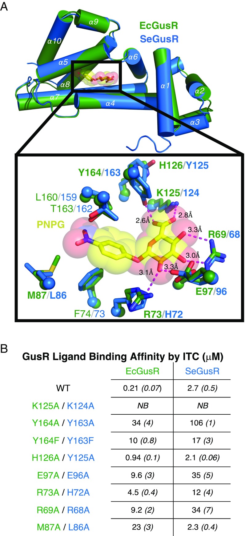
Fig. 4.
GusR effector-binding domain structure and function. (A) Superposition of PNPG (yellow)-bound EcGusR and SeGusR monomers (green and blue, respectively), with a close-up highlighting side chains that contact PNPG. Residues examined by mutagenesis are indicated (bold). (B) PNPG binding affinities measured by ITC for GusR wild-type (WT) and mutant GusR proteins. Error values are reported in parentheses and italicized, and represent SDs. NB, no binding.

