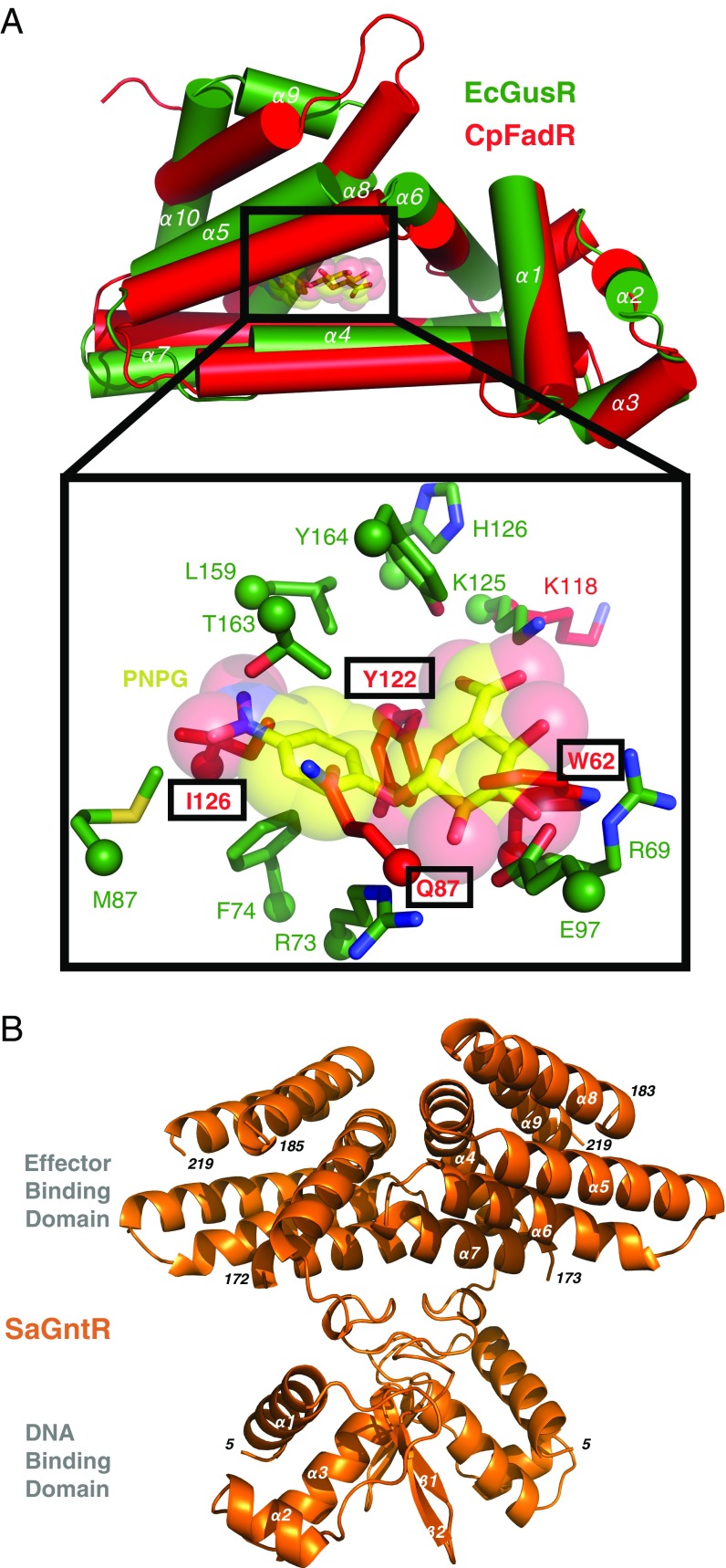
Fig. 6.
Putative GusR orthologs from C. perfringes and S. agalactiae. (A) Superposition of EcGusR and C. perfringes FadR monomers (green and red, respectively), with a close-up of residues of the putative effector-binding pockets. CpFadR side chains that sterically clash with PNPG are in bold and boxed. (B) Ribbon representation of the 1.9-Å crystal structure of the S. agalactiae GntR (orange; PDB ID code 6AZ6) homodimer.

