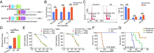
Fig. 2.
Molecular and functional characterization of the ZBTB16 (PLZF)-ABL1 fusion gene. (A) Structural and functional domains of wild-type ZBTB16, ABL1, and the representative fusion protein. Dashed lines indicated breakpoints of the wild-type proteins. (B) Cell proliferation of Jurkat cells transfected with ZBTB16-ABL1 (ZA) or vehicle (Control) seeded at a different density per well. (C) Cell-cycle distribution of Jurkat cells transfected with ZBTB16-ABL1 (ZA) or vehicle (Control). (D) Immunoprecipitated ABL1, BCR-ABL1 (BA), or ZBTB16-ABL1 (ZA) proteins were assayed for tyrosine kinase activity. The kinase activity of BA or ZA protein was compared with the activity of ABL1. (E) Viability of Jurkat cells transfected with ZBTB16-ABL1 (ZA) or vehicle (Control) upon exposure to dasatinib or imatinib. NR denotes “not reached.” (F) Kaplan–Meier survival analysis of ZBTB16-ABL1 (ZA) mice (n = 10) and control mice (n = 10). (G) Kaplan–Meier survival curves of control mice with ZBTB16-ABL1 (ZA) fusion (n = 6), imatinib (50 mg/kg) treated mice with ZA (n = 6), and dasatinib (5 mg/kg) treated mice with ZA (n = 6). Survival curves were compared by two-sided log-rank test. **P < 0.01; ***P < 0.001. Data are presented as mean ± SD from three independent experiments. Statistical significance was determined using two-sided Student’s t test.

