Fig. 2.
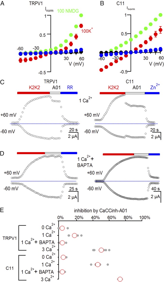
The C11 chimera is calcium permeable. (A) I–V relations for TRPV1-expressing cells before (black) and after application of 5 µM K2K2 in 100 mM K+ (red), 100 mM NMDG+ (green), or after application of 50 µM RR in 100 mM K+ (blue). Symbols are mean ± SEM for n = 5. (B) I–V relations for C11-expressing cells before (black) and after application of 5 µM K2K2 in 100 mM K+ (red), 100 mM NMDG+ (green), or after application of 2 mM Zn2+ in 100 mM K+ (blue). Symbols are mean ± SEM for n = 5. (C) Time course for activation of membrane currents following extracellular application of K2K2 and testing for inhibition by the calcium-activated chloride channel inhibitor CaCCinh-A01(A01) for cells expressing TRPV1 (Left) or the C11 chimera (Right). The external solution had 1 mM Ca2+. Cells were held at −60 mV, and voltage was stepped to +60 mV for 100 ms every 3 s. TRPV1 was subsequently inhibited by RR (50 µM; Left) and the C11 chimera by Zn2+ (2 mM; Right). The dotted horizontal line indicates the zero-current level. (D) Injecting cells with BAPTA eliminates the A01-dependent reduction in membrane current in the presence of 1 mM external Ca2+. (E) Summary of inhibition of K2K2-activated membrane current at +60 mV by 30 µM CaCCinh-A01 under different conditions. Data points for individual cells are shown as gray circles, and the mean ± SEM (n = 4–7) as red circles and black bars, respectively. The concentration of external Ca2+ is indicated in millimoles/liter.

