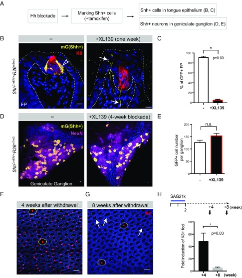
Fig. 7.
Hh antagonism causes loss of local Shh-producing cells in the taste buds. (A) Experimental design to determine the effect of prior Hh pathway blockade. (B) GFP staining of fungiform papillae from ShhCreER/+;R26mTmG mice. GFP+ (Shh-expressing) cells in the Shh-expressing basal cells marked in vehicle-treated group are denoted by open arrowheads. In XL139-treated group, GFP markings of Shh-producing neurons are denoted by arrows. (C) Quantified percentage of GFP-marked Shh-producing cells in fungiform papillae; tamoxifen was given after 2 wk of Hh blockade: 90.9 ± 2.81% vs. 5.3 ± 1.81%; P = 0.03; n = 4 mice in each group. (D) GFP+ (Shh-expressing) cells in the geniculate ganglion (mGFP in yellow; NeuN in magenta). (E) Quantification of GFP+ cell numbers per ganglion: 125.8 ± 9.8 vs. 153.3 ± 10.1; ns, not significant. Sample size, n = 12 and 7 ganglia from 6 and 4 mice in vehicle- and XL139-treated (4 wk), respectively. (F) Immunofluorescence image of tongue epithelium stained with K8 (red) and DAPI (blue). Normal TRC-containing fungiform papillae are denoted by yellow circles. (G) Residual ectopic K8+ foci after 4 wk are denoted by arrows. (H) Total number of K8+ foci normalized to the number of K8+ fungiform papillae quantified as 4 and 8 wk after removal of SAG21k treatment, normalized to untreated group (as 1, not plotted). n = 4 mice in each group (42 ± 11.7-fold; 4.5 ± 1.2-fold, P = 0.03). [Scale bars: 10 μm (B and D); 100 μm (F).]

