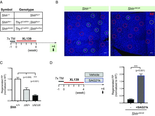
Fig. 8.
Shh from sensory neurons specifies the spatial patterning of TRC regeneration. (A) Genotype of mice used and experimental scheme to introduce neuronal Shh ablation before TRC regeneration. (B) Immunofluorescence images of tongue epithelia showing regenerated taste buds (green circles) in Shh+/+ and Shh∆N/∆N animals (partially regenerated taste buds denoted by yellow circles). (Scale bars, 100 μm.) Shown as composites of tile-scanned images stitched together by Zeiss ZEN software, as described in Materials and Methods. (C) Quantification of regenerated fungiform papillae as a function of Shh allele dosage, normalized to Shh+/+; n = 7, 5, and 15 mice in each group. (D) Experimental scheme to activate Hh pathway pharmacologically during TRC regeneration. Quantification of regenerated fungiform papillae in Shh∆N/∆N mice, analyzed after a 4-wk recovery period with either vehicle (DMSO) or SAG21k treatment. Shown as ratio normalized to Shh+/+ group in Fig. 6C. Number of mice, n = 15 and 11.

