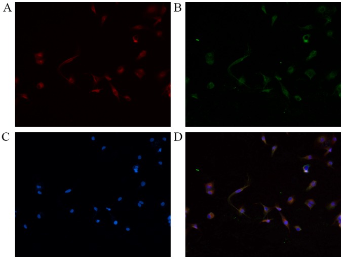
Figure 1.
Immunofluorescent double-staining of LX2 cells. (A) Red fluorescence indicates NRP-1 expression; (B) green fluorescence indicates PDGFR-β expression; (C) DAPI-stained nuclei. (D) Merge: Yellow fluorescence indicates co-expression of NRP-1 and PDGFR-β. Magnification, ×200. NRP-1, neuropilin-1; PDGFR-β, platelet-derived growth factor receptor-β.

