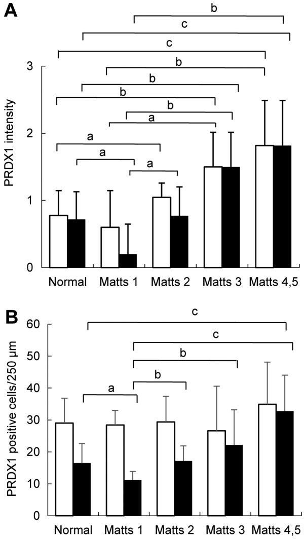
Figure 5.
PRDX1 expression in UC regenerative mucosa and normal mucosa. (A) Staining intensity of PRDX1 in epithelial cells of four stages: 0, 1, 2, and 3. (B) PRDX1-positive cells per 250 µm of stroma length were counted. Open bars, upper half of the crypt or lamina propria. black bars, lower half of the crypt or lamina propria. Data are presented as mean scores ± standard deviations. aP<0.05, bP<0.01, and cP<0.001.

