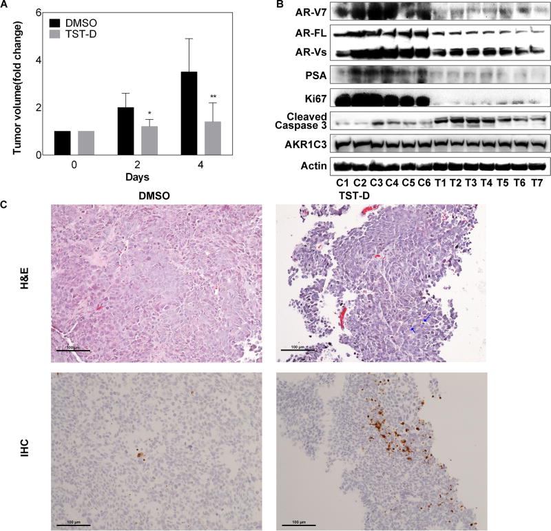
Figure 6. The in vivo effect of TST-D on CRPC xenograft.
(A) Mice bearing 22RV1 xenografts were treated with DMSO or 300 µg/kg/day TST-D; tumor volumes were measured every other day and tumor growth rate from each tumor was calculated based on pre-treatment. (B) The protein expression of AR-V7, AR-FL, PSA, Ki67, cleaved Caspase-3 and AKR1C3 from tumors collected 4 days after treatment was determined by western blot. C: control; T: treatment. (C) H&E staining was employed to examine drug-elicited cell death and confirmed by IHC staining using cleaved Caspase-3 antibodies (lower panel). Apoptic cells are marked with arrow. * P<0.05 ** P<0.01

