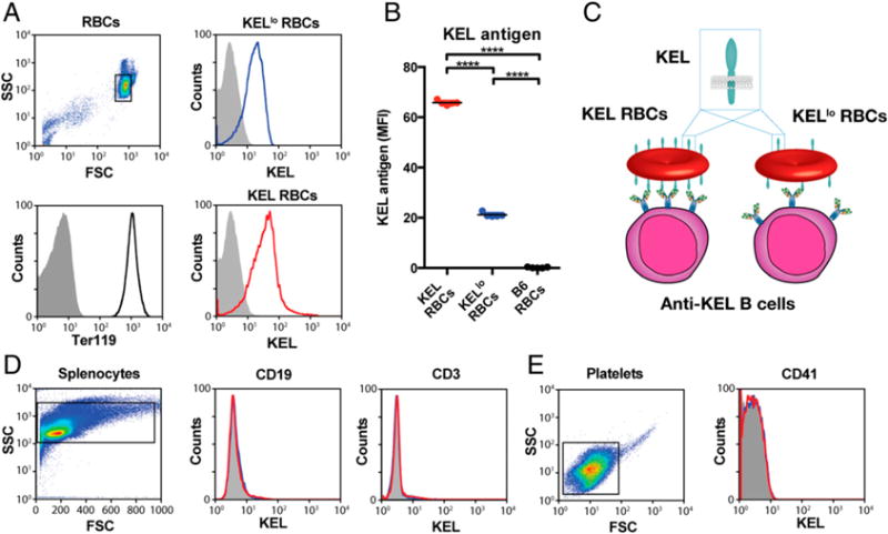
FIGURE 1.

Different founders stably produce RBCs with distinct levels of the KEL Ag. (A) Flow cytometric examination of KEL Ag expression on KELlo (blue) and KEL (red) RBCs. (B) Quantitative analysis of KEL Ag expression on KEL, KELlo, and C57BL/6 (B6) KEL− RBCs. (C) Schematic of potential differences in KEL and KELlo RBC interactions with the immune system. Analysis of KEL Ag expression on CD3+ T cells and CD19+ B cells (D) and CD41+ platelets (E) from KELlo (blue) and KEL (red) RBCs. Error bars indicate SEM. ****p < 0.0001, one-way ANOVA.
