
Figure 4. Choroid and Choriocapillaris Segmentation in a Patient With Intermediate Age-Related Macular Degeneration.
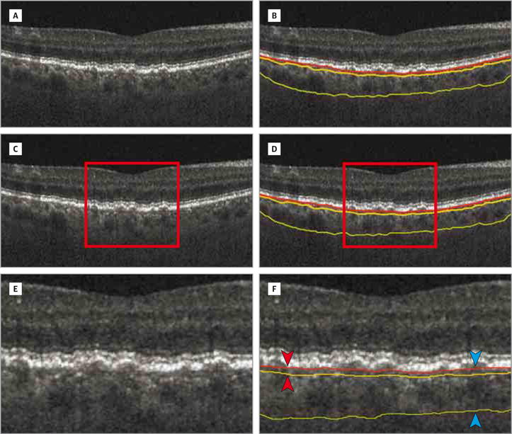
A, B-scan. B, B-scan with boundaries. B-scan with choriocapillaris-equivalent thickness (C and D). Enlarged views of parts C and D demonstrating choroidal vascular segmentation using the Bruch membrane as the top reference surface and the top of the choroidal vasculature as the bottom reference surface (E and F). F, The choriocapillaris-equivalent thickness is defined as the distance between the surfaces (red arrowheads). The choroidal thickness is defined as the distance between the Bruch membrane and choroidal posterior boundary (blue arrowheads). The red line indicates the Bruch membrane; yellow line, choroidal anterior boundary; and green line, choroidal posterior boundary.