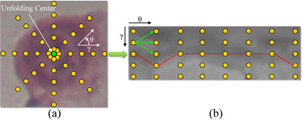
Fig. 2.

Schema of the graph-search based nucleus segmentation approach. Based on (a) the original cropped Cartesian image, (b) the graph is constructed from the image center (green point) using a polar transform. Yellow points represent pixels/nodes. Green arrows point to the successors of a node. (For interpretation of the references to color in this figure legend, the reader is referred to the web version of the article.)
