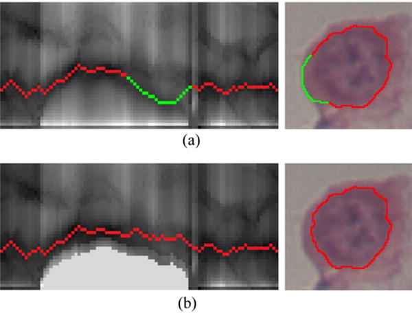
Fig. 4.

An example of context prior constraints, corresponding to the case in Fig. 3. (a) Incorrectly identified the cytoplasm-background border as the nucleus boundary, highlighted in green. (b) Correctly identified nucleus boundary after using context prior constraints. (For interpretation of the references to color in this figure legend, the reader is referred to the web version of the article.)
