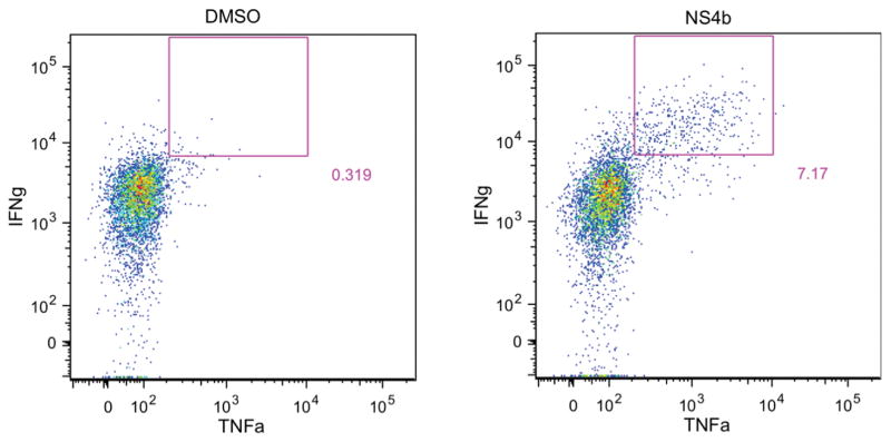
Figure 2.
Intracellular cytokine staining of brain cells 12d after WNV infection. C57BL/6 mice were infected s.c. in the footpad with 100pfu WNV-TX, and brain was harvested 12 days post-infection as previously described. Single cell suspensions were prepared and restimulated prior to staining for flow cytometry analysis. Cells are gated on CD8+ cells, and gate denotes IFNg+ TNFa+ cells after either DMSO (left) or peptide (right) stimulation.

