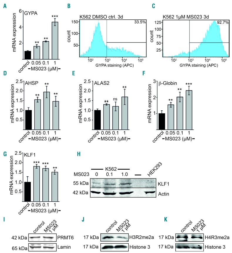
Figure 6.
Inhibition of PRMT6 increases erythroid gene expression. (A) GYPA expression increases at the mRNA level after treatment of K562 cells with the indicated concentrations of the PRMT6 inhibitor MS023 for 3 days. The control was treated with solvent only (DMSO). Expression was measured by quantitative reverse transcriptase PCR. (B,C) GYPA expression at the cell surface upon treatment of K562 cells with PRMT6 inhibitor for 3 days was determined by flow cytometry using an anti-CD235a-APC antibody. GYPA positivity is given in percent according to the indicated gating. (D–F) Expression of the erythroid genes AHSP, ALAS2 and β-globin increased upon treatment of K562 cells with the indicated concentrations of PRMT6 inhibitor for 3 days. (G,H) Expression of KLF1 increased upon inhibitor treatment on the mRNA and protein level. Expression was measured by quantitative reverse transcriptase PCR and western blot analysis. (I) Western blot analysis of PRMT6 protein expression upon inhibitor treatment of K562 cells for 3 days. (J) Western blot analysis of histone 3 methylation (H3R2me2a) upon inhibitor treatment of K562 cells for 3 days. (K) Western blot analysis of histone 4 methylation (H4R3me2a) upon inhibitor treatment of K562 cells for 3 days. Error bars indicate the standard deviation from four independent determinations. The P values were calculated using the Student t-test. *P<0 .05; **P<0.01; ***P<0.001.

