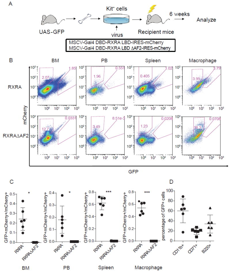
Figure 1. Natural RXRA ligands in mouse hematopoietic cells in vivo.

(A) Schema for bone marrow transplant procedure. Kit+ cells isolated from the bone marrow of UAS-GFP mice using MACS were transduced with one of the indicated retroviruses, then injected into lethally irradiated recipient mice. After 6 weeks of engraftment, recipient mice were sacrificed and their hematopoietic cells analyzed. (B) Representative FACS showing mCherry and GFP intensity in bone marrow cells (BM), peripheral blood (PB), spleen cells, and peritoneal macrophages from mice transplanted with UAS-GFP Kit+ bone marrow cells that were transduced with Gal4-RXRA or Gal4-RXRA-ΔAF2. (C) Ratio of GFP+mCherry+ cells relative to total mCherry+ cells in BM, PB, spleen, and peritoneal macrophages from mice transplanted with Kit+ bone marrow cells transduced with Gal4-RXRA (circles, N = 6 recipient mice) or RXRAΔAF2 (squares, N = 3 recipient mice). (D) The percentage of CD11b+ (circles), CD71+ (squares), and B220+ (triangles) cells in bone marrow GFP+mCherry+ from the recipient Gal4-RXRA mice (N = 6). Error bars represent standard deviation between individual mice. * P < 0.05, *** P < 0.001, T-test with Welch’s correction.
