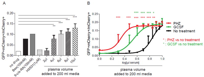
Figure 4. RXRA reporter activated by mouse plasma.

(A) UAS-GFP bone marrow Kit+ cells were transduced with Gal4-RXRA retrovirus and treated in culture with RXRA agonist (bexarotene and 9-cis-RA), antagonist (HX531), or mouse plasma as indicated, and the ratio of GFP+mCherry+ cells to total mCherry+ cells was determined. Cells were treated in a total volume of 200 μl media. Two way T-test compared against untreated control. (B) UAS-GFP bone marrow Kit+ cells were transduced with Gal4-RXRA retrovirus and treated in culture with plasma from indicated mice and the ratio of GFP+mCherry+ cells to total mCherry+ cells was determined. Error bars represent standard deviation between measurement of plasma obtained from individual mice (N = 3 mice per group). ANOVA with Tukey’s multiple comparisons compared results obtained at each plasma concentration. * P < 0.05, ** P < 0.01, *** P < 0.001.
