
Figure 1.
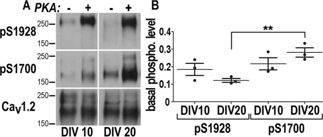
Quantification of Ser-1700 and Ser-1928 phosphorylation in cultured hippocampal neurons. Hippocampal neurons were extracted at 10 and 20 DIV, each sample was split into equal pairs, and CaV1.2 was immunoprecipitated. One sample of each pair but not the other was phosphorylated in vitro with purified PKA catalytic subunit under conditions that lead to near complete phosphorylation of the PKA site on CaV1.2, as quantified earlier (36). A, samples of sequential probing of immunoblots with antibodies against pSer-1928, pSer-1700, and finally total CaV1.2. B, immunosignals were quantified by film densitometry. pSer-1700 and pSer-1928 signals were corrected for differences in relative amounts of Cav1.2. Scatter plots show pSer-1700 and pSer-1928 levels detected in untreated samples as a fraction of the paired maximally phosphorylated samples. Of the Ser-1928 sites 18.4 ± 4.9% were phosphorylated in hippocampal cultures at 10 DIV and 12.1 ± 1.6% at 20 DIV. Of the Ser-1700 sites 21.6 ± 4.9% were phosphorylated at 10 DIV and 28.1 ± 3.8% at 20 DIV. All errors are given as S.D. Statistics: ANOVA: **, p = 0.03; n = 3 obtained in three independent experiments.