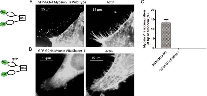
Figure 10.
Localization of myoVIIa-WT and myoVIIa-sh1 in HeLa cells. HeLa cells were transfected with pEGF-C1-myoVIIa-WT-GCN4 (A) or pEGFP-C1-myoVIIa-sh1-GCN4 (B) and then fixed with 4% paraformaldehyde and stained with 500 nm Texas Red phalloidin 14 h after transfection. C, percentage of cells with myoVIIa accumulation at the tip of filopodia. Left bar, pEGFP-C1-myosin VIIa WT-GCN4; right bar, pEGFP-C1-myosin VIIa shaker-1-GCN4. n = 3; mean ± S.E. (error bars).

