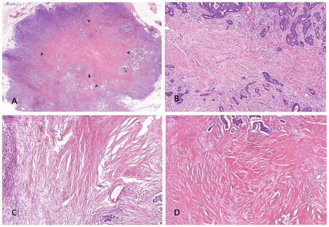
Figure 2.
Representative histology of fibrotic focus (FF) in invasive breast carcinoma. (A) Representative histology of FF; arrow heads indicate area of FF in invasive breast carcinoma [hematoxylin and eosin (H&E); magnification, ×10], (B) FF with fibrosis grade 1 showing fibroblastic proliferation in stroma with small amount of collagen (H&E; magnification, ×100), (C) FF with fibrosis grade 2 which intermediate between grade 1 and 3 fibrosis (H&E; magnification, ×100), (D) FF with fibrosis grade 3 showing mostly hyalinized collagenous stroma (H&E; magnification, ×100).

