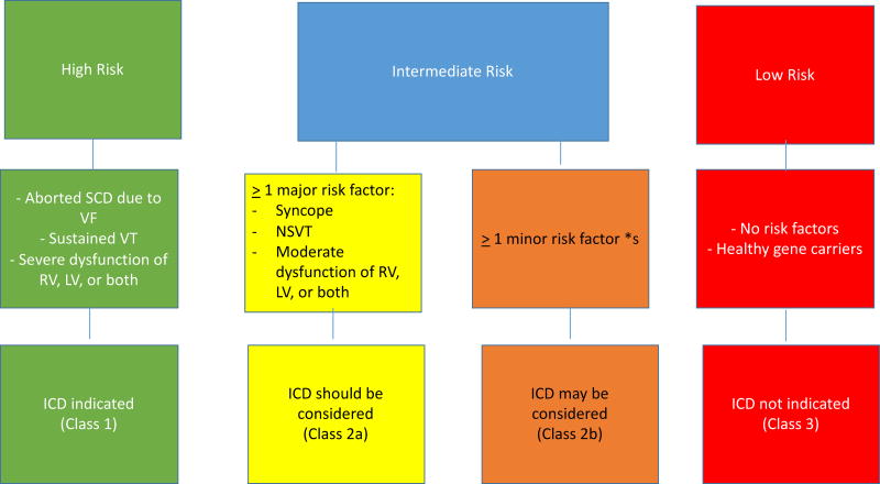
Figure 6.
Flow chart of risk stratification and indications to ICD implantation in ARVC/D. VT, ventricular tachycardia. RV, right ventricle. LV, left ventricle. Major risk factors include syncope, nonsustained VT, and moderate ventricular dysfunction, either RV (RVEF 36% – 46%, or LV (LVEF 36% – 45%). Minor risk factors* include: complex genotype, male gender, proband, positive EP study, T wave inversion in 2 of 3 inferior leads, T wave inversion ≥ 3 precordial leads. Modified from Corrado et al. (7).

