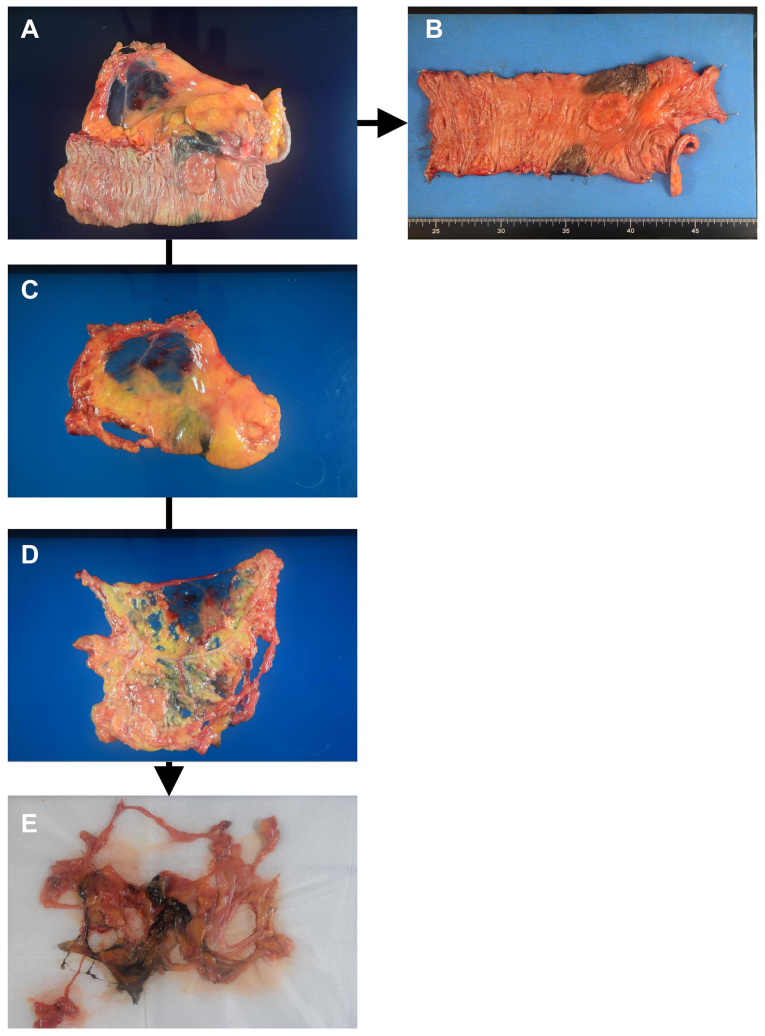
Figure 1.
Conventional procedure and fat dissolution technique. (A) The resected specimen is processed immediately after surgery. (B) The large bowel with colorectal cancer is pinned on a board. (C) The mesentery is dissected from the large bowel, and the serous membrane is cut with scissors and (D) the tumor-feeding arteries are exposed and the search for regional lymph nodes (LNs) is performed. (E) After fat dissolution, the tissue is transferred onto 5–10 sheets of gauze to drain the liquid, and the tissue is searched again for LNs for pathological examination.

