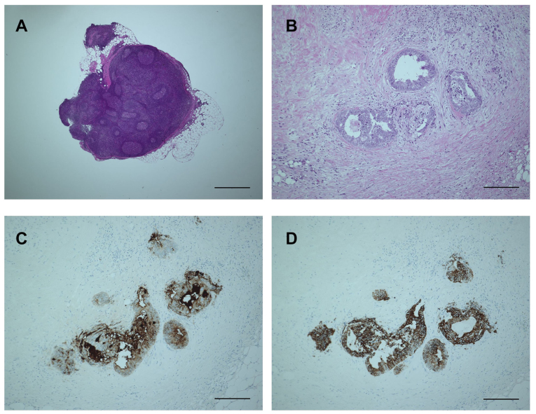
Figure 3.
Microscopic examination of obtained lymph node (LN) and cancer cells after fat dissolution. (A) The structure of the LN is well-maintained even after 60 min incubation in the fat dissolution liquid. The bar is 1,000 µm. (B) Hematoxylin and eosin staining of the tumor nodule is also not hampered by fat dissolution. The bar is 200 µm. (C and D) The tumor nodule is clearly positive for (C) carcinoembryonic antigen and (D) cytokeratin-20, compatible with the control results obtained at the first LN assessment (data not shown). The bars are 200 µm.

