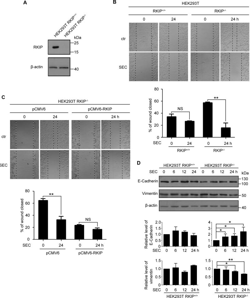
Fig. 1. SEC inhibited the cell migration of HEK 293T RKIP−/− cells.
(A) RKIP protein level in HEK293T RKIP+/+ and RKIP−/− cells. (B) A scratch on HEK293T RKIP+/+ and RKIP−/− cells was made, followed by incubation with SEC (20 µM) for 24 h. Relative wound closure was quantified by measuring the width of the wounds. (C) A scratch was made on HEK293T RKIP−/− cells transfected with pCMV6 empty vector and pCMV6-RKIP plasmid for 24 h, then treated with 20 µM SEC for 24 h. The width of the wounds was measured and relative wound closure was quantified. (D) HEK293T RKIP+/+ and RKIP−/− cells were treated with 20 µM SEC for 6, 12 and 24 h. The protein level of epithelial marker E-Cadherin and mesenchymal marker Vimentin was examined by western blot. Data are mean ± SEM; * p < 0.05, ** p < 0.01, NS p > 0.05, n = 3.

