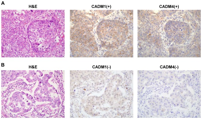
Figure 1.
Representative images of CADM1 and CADM4 immunohistochemistry staining with H&E staining in breast cancer tissue. (A) Tissue with positive CADM1 and CADM4 staining. (B) Tissue with negative CADM1 and CADM4 staining. Magnification, ×400. H&E, haematoxylin and eosin; CADM, cell adhesion molecule.

