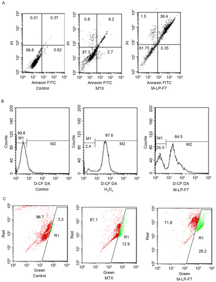
Figure 4.
(A) Cell apoptosis in HSC-3 cells after 48 h of treatment with MTX and M-LP-F7 at equivalent concentration of 70 µg/ml of MTX. (B) Pro-oxidant effect of MTX and M-LP-F7 in HSC-3 prostate cancer cells measured by intracellular ROS using a 2′,7′-dichlorofluorescin diacetate assay. (C) Intrinsic apoptotic pathway visible through the change from red to green fluorescence, marking the mitochondrial membrane potential change following treatment with MTX, M-LP-F7 or the control. FITC, fluorescein isothiocyanate; PI, propidium iodine; MTX, methotrexate; M-LP, MTX-entrapped liposomes.

