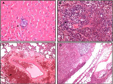
Fig. 2. Photomicrographs showing multifocal, acute degenerative tissue changes with the light blue staining indicating Gram-negative bacteria consistent with the pure isolation of P. multocida.

(A) Liver: necrosuppurative hepatitis, random with intralesional bacteria (arrow). (B) Spleen: splenitis, necrotizing with bacterial emboli. (C) Lung, perivascular hemorrhage (arrow) and edema. (D) Lymph node: lymphadenitis, necrosuppurative.
