. 2018 Jan 16;26:e20170084. doi: 10.1590/1678-7757-2017-0084

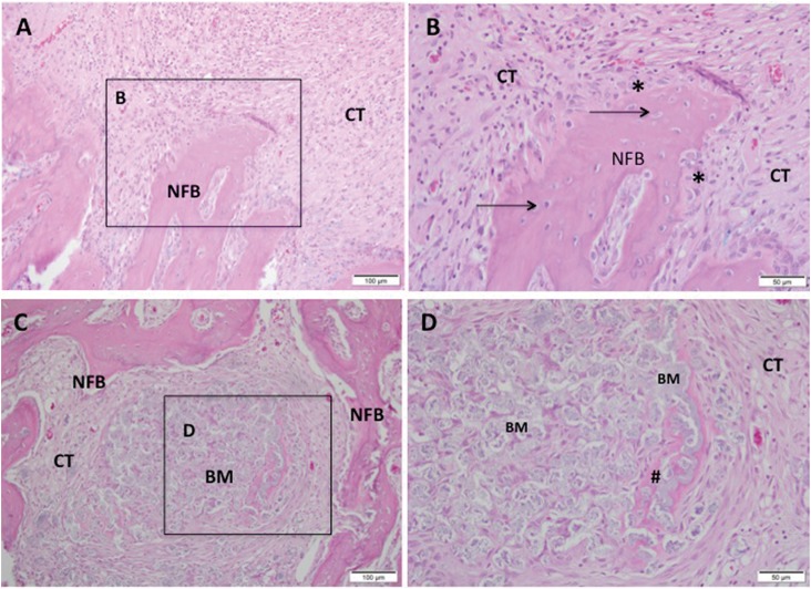
Figure 4. Representative photomicrographs of alveolar socket after 42 days. A and B: CHA group and C and D: SrCHA group. A and B: observe the presence of newly formed bone (NFB) with osteocytes (arrow) and osteoblastic paving (*) permeated by fibrocellular connective tissue (CT), with few inflammatory cells and absence of biomaterial (BM). C and D: presence of new-formed bone with connective tissue at the surroundings containing fragmented biomaterial and peripheric osteoid (#). A and C: 20-fold magnification and B and D: square with 40-fold magnification taken from A and C, respectively. BM: Biomaterial, NFB: Newly Formed Bone, CT: Connective Tissue; #: Osteoid, *: Osteoblasts. Hematoxylin and eosin stained. Bar: A and C: 100 μm; B and D: 50 μm.

Figure 4