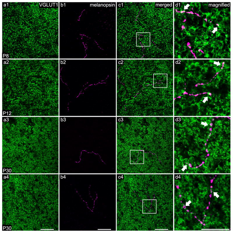
Figure 1. ORDs stratify in the OPL and lie closely opposed to cone photoreceptor terminals at all developmental ages and in the adult.
A single optical section (0.45 μm) generated from a series of confocal images masked to correct for the imperfect flatness of the retina. Immuno-positive photoreceptor terminals (VGLUT1) are labeled in green and melanopsin ORDs stratifying in the OPL are labeled in magenta. White areas (indicated with arrows) suggest regions of overlap between melanopsin staining and VGLUT1 staining. a1–d1) A branched ORD from a M1 melanopsin ganglion cell at P8. a2–d2) A branched ORD from a displaced M1 melanopsin ganglion cell at P12. a3–d3) An unbranched ORD from a M1 melanopsin ganglion cell at P30 (adult). a4–d4) A branched ORD from a displaced melanopsin ganglion cell at P30 (adult). Scale bar: 50 μm; Magnified scale bar: 25 μm.

