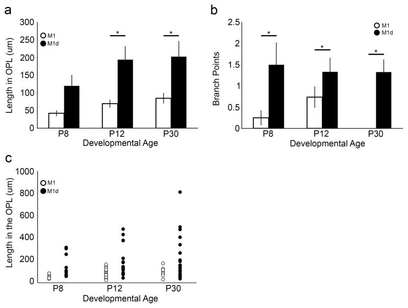
Figure 3. Melanopsin ganglion cell ORDs in the OPL form two morphologically distinct groups.
a) ORDs in the OPL that originate from M1 cells are significantly shorter than those that originate from M1d cells at P12 and P30 (n=5 retinas at each developmental age; p<0.05). b) ORDs in the OPL that originate from M1 cells have fewer branch points than those that originate from M1d cells (n=5 retinas at each developmental age; p<0.05). From these results, it appears that the greater length of M1d ORDs within the OPL is due to greater number of branch points. c) A scatter plot of ORD length in the OPL at different developmental time points. ORDs in the OPL from M1 cells have less dendrite in the OPL than those of M1d cells. ORDs that are longer (approximately >200 μm) have more than two branch points. Thus, the greater total length of M1 ORDs in the OPL seems to be due to a greater number of branch points. Error bars=SEM.

