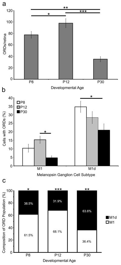
Figure 4. A subset of M1 and M1d melanopsin ganglion cells have ORDs in the INL and OPL during development and in the adult mouse retina.
a) The number of ORDs in the OPL and the INL changes throughout development. The number of ORDs increases from P8 to P12 (p<0.05) and then decreases from P12 to P30 (p<0.001). b) The percentage of M1 and M1d cells in the retina that have ORDs in the OPL and/or INL during development and in the adult. Unsurprisingly, the highest number of ORD-positive M1 and M1d cells is during development. The number of ORD-positive M1 cells decreases significantly from P12 to P30 (p<0.05), and the number of ORD-positive M1d cells decreases significantly from P8 to P30 (p<0.05). c) The percentage of ORDs in the OPL and INL that originate from M1 cells compared to those that originate from M1d cells. At P8 and P12, the majority of ORDs originate from M1 melanopsin ganglion cells (p<0.05 and p<0.001, respectively). However, at P30, the majority of ORDs originate from M1d melanopsin ganglion cells (p<0.01). Error bars=SEM.

